Corneal diseases are a group of rare, genetic eye disorders.
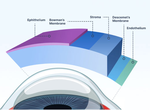
Corneal diseases are a group of rare, genetic eye disorders. With corneal diseases, abnormal material builds up in the cornea (the clear, front window of the eye). Most corneal diseases affect both eyes. They progress slowly and run in families.
The cornea has five layers:
Corneal diseases are caused by the build-up of material in one or more layers of the cornea. The material may cause the cornea to lose its transparency. This can cause loss of vision or blurred vision.
There are more than 20 different types of corneal diseases. They are generally grouped into three categories:
The symptoms of corneal disease depend upon the type of corneal dystrophy. Some people experience no symptoms. In others, the build-up of material in the cornea causes it to become opaque (not clear). This leads to blurred vision or vision loss.
If your ophthalmologist thinks you have a corneal dystrophy, they will examine your eye. They will also ask about your family history of eye disease.
Your ophthalmologist will use a slit lamp microscope to shine a thin, bright sheet of light into your eye. This helps the doctor examine the front part of your eye thoroughly.
If someone has no symptoms, a routine eye examination may show that they have corneal diseases. In some cases, genetic testing can identify corneal diseases.
Treatment for corneal diseases depends on:
If you do not have any symptoms, your ophthalmologist may monitor your eyes closely to see if the disorder is progressing. In other cases, eye drops, ointments or laser treatment may be appropriate.
A corneal abrasion is a scratch or scrape on the surface of your cornea.
Fingernails, makeup brushes, and tree branches are common culprits of corneal abrasions. Some other causes of corneal abrasion are rubbing your eye and having very dry eyes.

The cornea has many nerve cells. Cells called pain receptors transmit pain to tell us about possible damage to the eye’s surface. In fact, there are hundreds of times more pain receptors in our cornea than there are in our skin.
Your ophthalmologist will put dye called fluorescein on your eye’s surface. Then they will look at your cornea with an instrument called a slit lamp. The dye will highlight a cut or scratch on the cornea.
Your ophthalmologist will treat your eye based on what they find in the exam. Following are some options.
If your corneal abrasion is small, it probably will heal in 1–2 days. A larger corneal abrasion may take about a week to heal.

A cornea ulcer is an inflammation of the cornea — the clear, dome-shaped tissue on the front of your eye that covers the pupil and iris. Cornea ulcers may or may not be associated with an infection. Noninfectious cornea ulcers can be caused by a relatively minor injury, such as from wearing your contact lenses too long or getting a foreign body in the eye. Infectious cornea ulcers can be caused by bacteria, viruses, fungi, and parasites.
Causes of cornea ulcers include:

Symptoms of cornea ulcers include:
If you notice any of the symptoms of cornea ulcers or infection, make an appointment to see an eye specialist right away. Delays in diagnosis and treatment of cornea ulcers can lead to serious complications, including blindness.
Diagnosing cornea ulcers typically involve the following:
Treatment of noninfectious cornea ulcers vary depending on the severity. For example, with mild discomfort from a corneal scratch, artificial tear drops may be the only treatment. However, if a cornea ulcer is causing significant tearing and pain, topical eye medications may be necessary.
Treatment of infectious cornea ulcers vary depending on the cause of the infection.
Antibiotic eye drops are the primary treatment for bacterial cornea ulcers. Depending on the severity of the infection, drop frequency can range from around four times a day to every 30 minutes, even during the night. Sometimes oral antibiotics are used as a supplement.
Cornea ulcers caused by fungi typically require antifungal eye drops and oral antifungal medication.
If a virus is causing the infection, antiviral eye drops and oral antiviral medications may be effective. Other viruses need only supportive care such as artificial tear drops.
Cornea ulcers caused by the parasite acanthamoeba can be difficult to treat. Antiparasitic eye drops are used, but some acanthamoeba infections are resistant to medication and can require treatment for several months. Severe cases of acanthamoeba cornea ulcers may require a cornea transplant.
If a cornea ulcer doesn’t respond to medication, or if it causes permanent damage to the cornea that significantly impairs your vision, your eye care provider may recommend a cornea transplant.
Our eyes need tears to stay healthy and comfortable. If your eyes do not produce enough tears, it is called dry eye. Dry eye is also when your eyes do not make the right type of tears or tear film.

People tend to make fewer tears as they get older due to hormonal changes. Both men and women can get dry eye. However, it is more common in women—especially those who have gone through menopause.
Here are some other causes of dry eye:
Tell your ophthalmologist about all the prescription and non-prescription medicines you take.
Here are some of the symptoms of dry eye:
Having a lot of tears in your eyes with dry eye might sound odd. But your eyes make more tears when they are irritated by dry eye.
Your ophthalmologist will begin with an eye exam. They will look at your eyelids and the surface of the eye. They will also check how you blink.
There are many different tests that help diagnose dry eyes. Your ophthalmologist may do a test that measures the quality or the thickness of your tears. They may also measure how quickly you produce tears.
Your ophthalmologist might tell you to use artificial tears. These are eye drops that are like your own tears. You can use artificial tears as often as you need to. You can buy artificial tears without a prescription. There are many brands. Try a few until you find a brand that works best for you.
If you use artificial tears more than six times a day or are allergic to preservatives, you should use preservative-free tears. This is because the tears with preservatives may start to irritate your eyes.
Your ophthalmologist may suggest blocking your tear ducts. This makes your natural tears stay in your eyes longer. Tiny silicone or gel plugs (called punctal plugs) may be inserted in your tear ducts. These plugs can be removed later as needed. Your ophthalmologist could also recommend surgery that permanently closes your tear ducts.
Your ophthalmologist might have you use a prescription eye drop medication. This helps your eyes make more of their own tears.
If your eyes are irritated, your ophthalmologist can treat those problems. They may recommend:
Fuchs’ Dystrophy is a disease of the cornea. It is when cells in the corneal endothelium gradually die off. These cells normally pump fluid from the cornea to keep it clear. When they die, fluid builds up and the cornea gets swollen and puffy. Vision becomes cloudy or hazy.
Fuchs’ Dystrophy has two stages. In the early stage, vision is usually hazy in the morning. In the disease’s second stage, vision remains blurry all day.
People in their 30s and 40s may have Fuchs’ Dystrophy but not know it. Vision problems might not appear until age 50 or later. Women are more likely than men to have Fuchs’ Dystrophy. Family history of Fuchs’ Dystrophy also increases your risk of getting it.
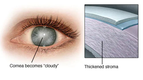
Normally, the cells lining the inside of the cornea (endothelial cells) help maintain a healthy balance of fluid within the cornea and prevent the cornea from swelling. But with Fuchs’ Dystrophy, the endothelial cells gradually die or do not work well, resulting in fluid buildup (edema) within the cornea. This causes corneal thickening and blurred vision.
Fuchs’ Dystrophy is usually inherited. The genetic basis of the disease is complex — family members can be affected to varying degrees or not at all.
In the early stage (stage 1) of Fuchs’ Dystrophy, you may notice few, if any, symptoms. Your vision may be hazy or blurry when you first wake up but improve throughout the day. This is because your eyes normally stay moist when they are closed during sleep. But when you are awake, the fluid dries normally.
In the later stage (stage 2), your blurry or hazy vision will not get better as the day goes on. Too much fluid builds up during sleep and not enough dries up during the day. Also, tiny blisters may form in the cornea. The blisters get bigger and eventually break open, causing eye pain. Here are other symptoms:
Your ophthalmologist will look closely at your cornea and measure its thickness. This is called pachymetry. They will also check for tiny blisters on the front surface of the cornea and drop-like bumps of the back surface of the cornea (guttae). Using a special photograph of your cornea, your ophthalmologist may count your endothelial cells.
There is no cure for Fuchs’ Dystrophy. However, you can control vision problems from corneal swelling. Your treatment depends on how Fuchs’ Dystrophy affects your eye’s cells.
Here are treatments for early Fuchs’ Dystrophy:
For very poor vision or scarred corneas, you may need a corneal transplant. This surgery could be one of three types:
Your ophthalmologist will discuss what treatments are best for your condition.
Doctors do not know for sure why people have keratoconus. In some cases, it appears to be genetic (passed down in families). About 1 out of 10 people with keratoconus have a parent who has it too. Keratoconus is also associated with:
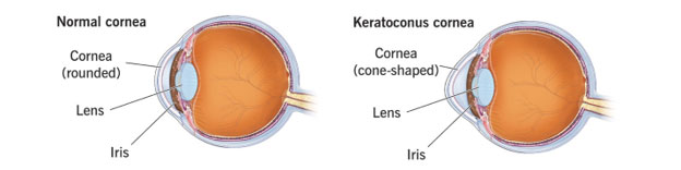
Keratoconus often starts when people are in their late teens to early 20s. The vision symptoms slowly get worse over a period of about 10 to 20 years.
Keratoconus often affects both eyes, and can lead to very different vision between the two eyes. Symptoms can differ in each eye, and they can change over time.
In the early stage, keratoconus symptoms can include:
In later stages, keratoconus symptoms often include:
Keratoconus usually takes years to go from early- to late-stage. For some people, though, keratoconus can get worse quickly. The cornea can swell suddenly and start to scar. When the cornea has scar tissue, it loses its smoothness and becomes less clear. As a result, vision grows even more distorted and blurry.
A routine eye exam may cause a suspicion of keratoconus, but it is best diagnosed by special scans of your cornea. Your optometrist or ophthalmologist will review these scans for information about thickness and curvature to diagnose keratoconus and monitor for progression.
Keratoconus treatment depends on your symptoms. When your symptoms are mild, your vision can be corrected with eyeglasses. Later you may need to wear special hard contact lenses to help keep vision in proper focus.
Here are other ways that your ophthalmologist might treat keratoconus:
With keratoconus, try to avoid rubbing your eyes. This can damage thin corneal tissue and make your symptoms worse.
If you have itchy eyes that cause you to rub, speak to your ophthalmologist about medicines to control your allergies.
Pterygium is a growth of fleshy tissue (has blood vessels) that may start as a pinguecula. It can remain small or grow large enough to cover part of the cornea. When this happens, it can affect your vision.

Pterygium is believed to be caused by a combination of exposure to ultraviolet (UV) light from the sun, wind, and dust.
Symptoms of pterygium can range from mild to severe. They include:
The diagnosis is made by slit-lamp examination of the wing-shaped limbal growth at the characteristic location within the palpebral fissure. The diagnosis is most often clear clinically, but histopathologic confirmation is performed routinely, as there can be associated dysplasia of the overlying tissue.
In many cases pterygium does not need to be treated. However, if your eyes are uncomfortable or your vision is affected, you may need treatment.
Your ophthalmologist can treat the discomfort, redness, or swelling from a pterygium with lubricating or steroid eye drops.
If a pterygium grows large enough to cause problems, your ophthalmologist will probably recommend surgery to remove it. Once the pterygium is removed, your surgeon may transplant a thin piece of normal tissue onto the affected area. This technique helps reduce the chance that your pterygium will grow back.
The best way to keep pterygium from coming back is to avoid sunlight, dryness, and dust.