The retina is the layer of cells that line the back of the inner wall of your eye.
The retina is the layer of cells that line the back of the inner wall of your eye. Our retinas sense light and send signals to our brain to see. Many different eye disorders affect the retina. Retinal diseases are detected during your annual comprehensive eye exam.


One treatment, intravitreal injections with anti-VEGF medications (Lucentis, Avastin, or Eylea), helps to reduce the growth of abnormal blood vessels, slow their leakage, and slow vision loss. The injections treat several retina diseases, including diabetic retinopathy, macular degeneration, and retinal vein occlusion.

Vitrectomy surgery is a specialized procedure (done off-site via referral) to treat severe eye injuries, diabetic retinopathy, retinal detachments, macular puckers (wrinkling of the retina), and macular holes.
During a vitrectomy operation, the surgeon makes tiny incisions in the sclera (the white part of the eye). Using a microscope to look inside the eye and microsurgical instruments, the surgeon removes the vitreous and repairs the retina through the tiny incisions. Repairs include removing scar tissue or a foreign object if present.
The retina is treated with a laser to reduce future bleeding or fix a retina tear during the procedure. An air or gas bubble that slowly disappears on its own may be placed in the eye to help the retina remain in its proper position, or a special fluid may be injected into the vitreous cavity (and removed later).
Recovering from vitrectomy surgery may be uncomfortable, but the procedure often improves or stabilizes vision. Once the blood- or debris-clouded vitreous is removed and replaced with a transparent medium (usually a saltwater solution), light rays can once again focus on the retina. Vision after surgery depends on how damaged the retina was before surgery.

Patients with diabetes may develop diabetic retinopathy (when the tiny blood vessels in your retina weaken).
There are two main types of diabetic retinopathy. Nonproliferative retinopathy is when your retina blood vessels leak and then close. In the early stages, there are few symptoms, but as it progresses, fluid deposits and swelling can cause moderate to severe vision loss.
Proliferative diabetic retinopathy (PDR) happens when the retinal blood vessels are so damaged that they close off. In response, the retina grows new, fragile blood vessels. Unfortunately, these new blood vessels are abnormal. They multiply on the retina’s surface and do not resupply it with blood.
Occasionally, these new blood vessels leak and cause a vitreous hemorrhage. Blood in the vitreous, the clear gel-like substance that fills the inside of the eye, blocks light rays from reaching the retina. A small amount of blood will cause dark floaters, while a significant hemorrhage might block all vision, leaving only light and dark perception. The new blood vessels can also cause scar tissue to grow. The scar tissue shrinks, wrinkling and pulling on the retina and distorting vision. The macula may detach from its normal position and cause vision loss if the pulling is severe.
Diabetic retinopathy develops slowly, often with no early warning signs. If you have diabetes, you should be seen by your Ophthalmologist regularly and have a dilated eye exam at least once a year. Keeping your blood sugar, blood pressure, and cholesterol under control is essential. In later stages of the disease, injections or surgery may be required to treat it.
Treatments include intravitreal injections and laser surgery to shrink the abnormal blood vessels and reduce the risk of bleeding. A vitrectomy is performed if a vitreous hemorrhage is not cleared up or a retinal detachment is detected.
Macular degeneration is damage or breakdown of the macula, the small part of the retina responsible for central vision. It affects both distance and close vision and can make some activities like threading a needle or reading very difficult or impossible. Macular degeneration is the leading cause of severe visual loss in people over 65.
Age-related macular degeneration (AMD) is one of the most common causes of poor vision after age 60. Although the specific cause is unknown, age is the most significant risk factor, heredity, blue eyes, high blood pressure, cardiovascular disease, and smoking. AMD accounts for 90 percent of new legal blindness in the US.

The visual symptoms of AMD involve loss of central vision. While peripheral vision is unaffected, one loses the sharp, straight-ahead vision necessary to drive, read, recognize faces, and generally look at details. Imagine seeing a clock on the wall but unable to make out the time or unable to read because you could not see words on the page.
There are two types of AMD. Nine out of 10 people have atrophic (dry) AMD, which results in the thinning of the macula, the area of the retina responsible for central vision. Dry AMD takes many years to develop. Currently, there is no treatment. But there have been studies conducted by the National Eye Institute on age-related eye diseases that found some vitamin supplements to help delay and prevent dry AMD from advancing to wet AMD. As with any vitamin regimen, it is essential to consult your physician first.
Exudative (wet) AMD occurs when abnormal blood vessels grow underneath the retina. These unhealthy vessels leak blood and fluid, which can scar the macula. In-office intravitreal injections with anti-VEGF can help slow vision loss.
There is promising AMD research. In the meantime, high-intensity reading lamps, magnifiers, and other low-vision aids help people with AMD make the most of their remaining vision. Laser surgery may also be used to help prevent further vision loss.

A detached or torn retina happens when the retina lifts away from the back of your eye like wallpaper peeling off a wall. As you get older, the vitreous, the clear gel-like substance that fills the inside of your eye, tends to shrink slightly and take on a waterier consistency. Sometimes as the vitreous shrinks, it exerts enough force on the retina to make it tear. They are caused by age, heredity, nearsightedness, cataracts, glaucoma, and severe eye injuries.
The appearance of flashing lights, floating objects, or a gray curtain moving across the field of vision is all indications of a retinal detachment. If any of these occur, see an ophthalmologist right away.
A retinal detachment is a severe problem that almost always causes blindness unless treated.
If the retina is detached, it must be reattached before sealing the retinal tear. There are three ways to repair retinal detachments.
Pneumatic retinopexy involves injecting a special gas bubble into the eye that pushes on the retina to seal the tear.
The scleral buckle procedure drains the fluid from under the retina before a flexible piece of silicone is sewn on the outer eyewall to support the tear while it heals.
Vitrectomy surgery removes the vitreous gel from the eye, replacing it with a gas bubble, slowly replaced by the body’s fluids.

The macula is the part of the retina responsible for acute central vision, the vision one uses for reading, watching television, and recognizing faces. A macular hole is a small round opening in the macula. The hole causes a blind spot or blurred area directly in the center of your vision.
Most macular holes occur in the elderly. When the vitreous (the gel-like substance inside the eye) ages and shrinks, it can pull on the thin tissue of the macula, causing a tear that can eventually form a small hole. Sometimes injury or long-term swelling can cause a macular hole. No specific medical problem is known to cause macular holes.
Vitrectomy surgery, the only treatment for a macular hole, removes the vitreous gel and scar tissue pulling on the macula and keeping the hole open. The eye is then filled with a special air bubble to push against the macula and close the hole. The air bubble will gradually dissolve, but the patient must maintain a face-down position (in a special massage-style chair) for one to two weeks to keep the gas bubble in contact with the macula. The success of the surgery often depends on how well the position is maintained.
With treatment, most macular holes shrink, and some of the lost central vision slowly returns. The amount of visual improvement typically depends on the length of time the hole was present. Some people with normal vision in the other eye may not want surgery since vitrectomy surgery cannot completely restore vision.
Floaters are specks or clouds that move into your field of vision as you look at a blank wall or a clear blue sky. Most people have floaters but do not notice them until they become numerous or more prominent. In most cases, floaters are part of the natural aging process. They look like cobwebs, squiggly lines, or floating bugs and appear to be in front of the eye but are floating inside. Floaters can get in the way of clear vision, often when reading. Try looking up and then down to move the floaters out of the way. While some floaters may remain, many of them will fade over time.

The appearance of flashing lights (flashers) comes from the traction of the vitreous gel on the retina at the time of vitreous separation. Flashes look like twinkles or lightning streaks. You may have experienced the same sensation if you have ever been hit in the eye and seen stars.
Floaters and flashes are sometimes associated with retinal tears. When the vitreous shrinks, it can pull on the retina and cause a tear. If new floaters appear suddenly or you see sudden flashes of light, see an ophthalmologist immediately.
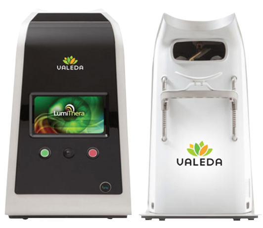
If you’ve been told there’s nothing you can do about dry AMD, there’s new hope. Valeda Light Therapy is an FDA-approved treatment that works differently than other options. Instead of just slowing progression, it’s designed to stimulate your retinal cells and help improve your vision. Quick sessions. Minimal side effects.
Valeda delivers a non-invasive, multiwavelength, light-based therapy that utilizes photobiomodulation (PBM) to treat dry age-related macular degeneration (AMD). This targeted treatment delivers specific wavelengths of light to the retina, stimulating cellular activity to improve vision and slow disease progression. Clinical studies have shown that Valeda can improve visual outcomes. Here’s the science:
Your retina has cells called photoreceptors. In dry AMD, these cells gradually break down, causing vision loss. Valeda’s light therapy is designed to energize these cells at the mitochondrial level, essentially giving them the boost they need to function better.
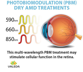
The result? Your retina may work more efficiently, potentially improving your vision and reducing drusen (the protein deposits that accumulate in dry AMD).
What makes it different:
At the initial assessment, a routine retinal examination with imaging is performed to verify that you are suitable for treatment. Follow-up retinal examinations with imaging will occur following your last treatment in each cycle.
The treatment is safe, non-invasive, and generally pain free. There is no down-time required following treatment.
A follow-up retinal examination with imaging is performed after your last treatment in each cycle.

Session details:
Treatment schedule:
The diagnosis is made by slit-lamp examination of the wing-shaped limbal growth at the characteristic location within the palpebral fissure. The diagnosis is most often clear clinically, but histopathologic confirmation is performed routinely, as there can be associated dysplasia of the overlying tissue.
In many cases pterygium does not need to be treated. However, if your eyes are uncomfortable or your vision is affected, you may need treatment.
Your ophthalmologist can treat the discomfort, redness, or swelling from a pterygium with lubricating or steroid eye drops.
If a pterygium grows large enough to cause problems, your ophthalmologist will probably recommend surgery to remove it. Once the pterygium is removed, your surgeon may transplant a thin piece of normal tissue onto the affected area. This technique helps reduce the chance that your pterygium will grow back.
The best way to keep pterygium from coming back is to avoid sunlight, dryness, and dust.